Thursday, April 11, 2019

School Board/Position Paper: Physical Activity in Adolescents

by Lauren Graf, Neuroscience Major at Kenyon College, Class of 2020
I am a junior neuroscience major at Kenyon, and I am taking a class on the Behavioral Neuroscience of Adolescence. One of the requirements of the class is to present the neuroscientific findings of a topic of interest to the community. I chose to focus on physical activity in children and adolescents, which I then presented information about to the Knox County School Board. 


Children and adolescents are not getting the amount of physical activity needed to maintain a healthy lifestyle. Exercising helps to keep both our body and our brains healthy and functioning at high levels. The CDC recommends that students in elementary through high school have at least a 20 minute recess, physical education, and physical activity in the classroom each day. School takes up a lot of adolescents’ time during the day, and it primarily requires them to remain seated for a large portion of that time. However, physical activity may actually improve children’s performance in school. I will discuss the neuroscientific findings behind this and the ways in which we could improve children’s amount of physical activity during school. 
Students who achieve higher fitness levels also achieve higher scores academically. Chomitz of Tufts University and her colleagues tested children in Cambridge Public Schools in fourth through eighth grade. They compared their fitness level, as determined by passing 5 fitness tests, to their academic achievement, which was measured by MCAS Mathematics and English scores (Chomitz et al., 2009). The researchers concluded that there was a significant positive relationship between number of fitness tests passed and achievement on the exams. The more fitness tests passed, the better the students did on the exams. How might physical activity have a positive impact on increased cognitive function? First, executive functioning refers to the higher-level cognitive processing associated with goal-directed behavior, which can include inhibition, flexibility, and working memory. The prefrontal cortex, or PFC, is an important area associated with executive functioning. Tuckmann and colleagues provided evidence that children in elementary and middle school who participated in running for 30 minutes per day, 3 to 5 days per week, had increased improvement in creativity and flexibility based on improvements in the Alternate Uses Test and the Torrance Test of Creative Thinking. Davis et al. had children participate in aerobic games, such as running games or basketball, for 20 or 40 minutes, and then measured their executive functioning capabilities as compared to a control group. The results were improved performance in executive functioning tasks as measured by the Cognitive Assessment System, along with an increase in mathematic achievements and increased PFC activation for children who participated in the games.
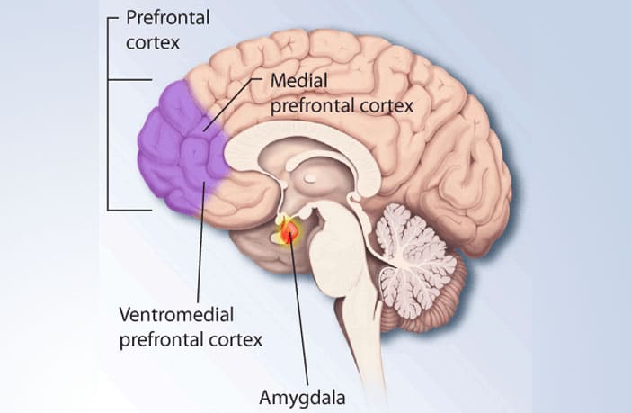
Source: https://www.psypost.org/2017/06/depressed-people-medial-prefrontal-cortex-exerts-control-parts-brain-49168
Not only does exercise help to improve higher-level cognitive functioning, but studies from Holmes and van Praag have shown that aerobic exercise increases regulation of growth factors in the brain, including the brain-derived neurotropic factor, or BDNF, which is important in the neurogenesis, or growth of brain cells, particularly in the hippocampus. The hippocampus is an area of the brain associated with memory, so when there are greater amounts of BDNF in the brain, there are more hippocampal cells which can help to facilitate learning and memory.
Source: https://www.epilepsyresearch.org.uk/the-hippocampus-what-is-it/
Based on these findings, it is evident that at least 30 minutes of physical activity is crucial for children, and can help them to attain high academic achievements. Schools must find more time and ways to integrate physical activity into the school day, as well as promote active play activities outside of school. These can include creating lesson plans that get them outside, playing a game that involves some sort of physical activity, and continuing physical education year-round, all the way through high school. A new athletic facility is being built for the community, which would also be a great resource for adolescents to use. These are all ways that can get students moving which leads to optimal brain function and increased academic performance.


References
Abdelbary, M. (2017). Learning in Motion: Bring Movement Back to the Classroom. Teacher Education Week.

Adolescent and School Health | CDC. (n.d.). 

Best, J. R. (2010). Effects of Physical Activity on Children's Executive Function: Contributions of Experimental Research on Aerobic Exercise. Developmental review : DR30(4), 331-551.

Chomitz, V. R., Slining, M. M., McGowan, R. J., Mitchell, S. E., Dawson, G. F., Hacker, K. A. (2009). Is there a relationship between physical fitness and academic achievement Positive results from public school children in the northeastern United States. Journal of School Health.79, 30-37.

Holmes, P. V. (2006). Current findings in neurobiological systems' response to exercise. In: L. Poon, W. Chodzo-Zajko, P. D. Tomporowski (Eds.), Active living, cognitive functioning, and aging (pp. 75–89). Champaign, IL: Human Kinetics.

van Praag, H. (2006). Exercise, neurogenesis, and learning in rodents. In: E. O. Acevedo & P. Ekkekakis (Eds.), Psychobiology of physical activity (pp. 61–74). Champaign, IL: Human Kinetics.

Wednesday, April 10, 2019

The Biological, Social, and Behavioral Changes Associated with Adolescence

By Rose Fisher, Undecided, '22

This is an annotated bibliography of an article by Sarah-Jayne Blakemore and Kathryn Mills named: Is Adolescence a Sensitive Period for Sociocultural Processing? I chose this article because I think it provides a good overview of the biological, neurological, social, and behavioral changes associated with adolescence.

Blakemore, S. J., & Mills, K. L. (2013). Is Adolescence a Sensitive Period for Sociocultural Processing? Annual Review of Psychology Vol. 65:187-207, Retrieved March 28, 2019, from https://www.annualreviews.org/doi/10.1146/annurev-psych-010213-115202


In this article, authors Blakemore and Mills review both the functional and structural changes that arise in the human brain during adolescence, examining other experiments and studies to provide evidence for their claims. The authors explain that adolescence is marked both by puberty and by social and behavioral changes, and that during adolescence, teenagers become increasingly interested in socialization and peer-acceptance (Blakemore & Mills, 2013). Blakemore and Mills address how these changes can lead to mental health disorders, as adolescents’ interactions with and perceptions of others and themselves rapidly change, and as the neurochemistry of the adolescent brain evolves. Specifically, the authors note how the development of the HPA axis, combined with increased levels of glucocorticoids during adolescence can contribute to psychological disorders. Blakemore and Mills also discuss the development of a number of social cognitive processes, including face processing and biological motion detection, which undergo significant development during adolescence. Face-processing is  especially important for adolescent development, as it is a crucial tool for reading and understanding the emotions of others. There are, however, other more complicated social processes that involve interpreting other peoples’ emotions and thoughts and making difficult personal decisions. These processes are harder to evaluate but are extremely important to adolescent growth. The neurochemical changes occurring in the adolescent brain also allow teenagers to develop deeper social emotions. The authors define social emotions as those which result from understanding other’s opinions and perceptions. Guilt, embarrassment and pride are provided as examples.
Blakemore and Mills explain how the biological social brain network, which consists of the “dorsal medial prefrontal cortex (dmPFC), temporoparietal junction (TPJ), posterior superior temporal sulcus (pSTS), and anterior temporal cortex (ATC)”, develops during adolescence (Blakemore & Mills, 2013, p.192). The social brain network is needed to perceive others’ emotions and thoughts, and to understand one’s own emotions. The authors discuss how changes in the structure of the brain, namely in the thickness, surface area, and concentration of white and grey matter in the cerebral cortex, generate changes in social processing. Lastly, Blakemore and Mills explain that because the prefrontal cortex is still developing during adolescence, adolescents are less able to plan ahead, make good decisions, and understand the consequences of their actions. Because they are still developing these skills, adolescents are thus more prone to engage in risky behavior. The authors note that individuals who have negative interactions with their environments as adolescents are significantly more likely to engage in risky behavior as adults, and advocate that abrupt changes in schooling, social settings, or home-life can negatively affect adolescent growth.


Image result for The HPA axis
The HPA Axis
https://neuroscientificallychallenged.com/blog/2014/5/31/what-is-the-hpa-axis


Image result for the biological social brain network
The Structure of the Biological Social Brain Network
https://www.nature.com/articles/nrn2353


A Review of Two Brain Research Tools

by Andres Tuccillo, Kenyon College, '22  

This is a listing of two commonly used neurological research tools (the fMRI and EEG). As well as a common explanation of their functions, the numerous strengths and weaknesses of each research tool will be explained below. 

One very popular and useful research tools is the fMRI (or functional magnetic resonance imaging). This technique is similar to the MRI but it allows researchers to see the brain, for lack of better terms, “in motion.” Basically, researchers are able to see which regions of the brain are activated when certain stimuli are encountered or when a certain cognitive operation is needed. In research specifically angled toward adolescence, fMRI has been useful to see how brain functions differ between age groups. Furthermore, fMRIs are able to take these images because of the simple fact that when a particular part of the brain is used, blood flow to that region aa well as blood oxygenation increases. This is commonly known as hemodynamics. Oxygenated red blood cells carries oxygen to the brain, displacing deoxygenated red blood cells. Oxygen is carried in red blood cells by the molecule hemoglobin. Deoxygenated hemoglobin (dHb) is more magnetic than oxygenated hemoglobin (Hb). The different magnetic properties of oxygenated and deoxygenated blood can be visualized by the fMRI. There are many strengths attributed to the fMRI. First off, it is a noninvasive procedure. Subjects are not poked and prodded; there are no outside elements invading their body. The fMRI is able to examine living, healthy, and developing brains by taking detailed images. However, there are limitations. These include that fact that subjects need to be motionless for the procedure, and this is not guaranteed, especially with adolescence. Also, a subject may be claustrophobic, so the tight space of the fMRI machine would be terrifying. Lastly, as of right now, there is no consensus as to how exactly analyze fMRI data. 

Another brain research tool is the electroencephalogram (EEG). An EEG measures the electrical impulses passed between neurons. To do this, the EEG test uses electrodes (consisting of small metal discs with thin wires) placed on the scalp. These electrodes collect the electrical information from the firing neurons, amplify them and it is this information that appears on a computer screen. Furthermore, neurons communicate through five different waveforms: alpha, beta, gamma, theta, and delta waves. Each different waveform signals a different cognitive process. This is how EEGs are able to tell what a person is thinking/doing and which regions are activated. Alpha waves signal that someone is waking up from sleep and beginning cognitive function; baeta waves signal anxiety, depression, or the use of sedatives; gamma waves signal peak focus and consciousness; theta waves are most common in children and young adults; and delta waves occur in young children during sleep. One major strength of the EEG is that it is a vert safe procedure. The electrodes do not produce any sensation when used, they merely collect information. Also, the EEG is less sensitive to participants moving than the MRI. Finally, the EEG records information in real time. This gives an extremely accurate image of the region of the brain that is being studied. The main limitation is that it does not provide a detailed image of the brain. Its spatial resolution is poor at best because only impulses from the surface of the brain are acquired.


Image collected from a fMRI:

Image result for fmri 
fMRI_brain-scan.jpg

Digram of an EEG: 
Image result for eeg
ZrmxJRu.jpg

Reference List:
Galván, A. (2017). The Neuroscience of Adolescence. New York, NY: Cambridge University Press.

Adolescent Risk Taking

by Kylie Baker-Williams, Kenyon College, 2022

This is an annotated bibliography of risk taking in adolescents. Here I will review three articles that look at why it happens, who's at risk, and how peers influence risk taking behaviors.

So, what is happening in the brain that results in this risky behavior? Qu, Y., Galván, A., Fuligni, A. J., Lieberman, M. D., & Telzer, E. H. (2015). Longitudinal changes in prefrontal cortex activation underlie declines in adolescent risk taking. Journal of Neuroscience, 35, 11308-11314.
A study by Galván, Fuligni, Lieberman, and Telzer was done in order to determine the way the prefrontal cortex functions in the context of adolescent risk-taking. The researchers took a group of 24 adolescents and gave them two fMRI scans, about a year and a half apart. At both scans, the participants reported their involvement in risk-taking activities using the Youth Self-Report. In order to examine the participants' sensitivity to risky behaviors, they completed the Balloon Analog Risk Task while undergoing the fMRIs. The final results from this study do not include two of the participants due to excessive head movement. The researchers did not find an overall change in behavior with the BART test, however, declines in risk-taking on the BART did covary with declined self-reports of risky behavior. When they looked at the neural results they found heightened activation in cognitive control and reward-related networks during the first and second scan. When they compared the two scans they found that the second scan, the one taken about 1.5 years later, showed less activation of the VLPFC in the participants compared to their first scans. They also found that the participants that reported less risk-taking behavior also had less activation in their VLPFC and VS than other participants. These findings suggest that higher activation of the VLPFC is an important part of risk-taking behavior in adolescents, and as we get older the region becomes less sensitive and active.

What else is going on? Galvan, A., Hare, T., Voss, H., Glover, G., & Casey, B. (2007). Risk-taking and the adolescent brain: Who is at risk? Developmental Science, 10.
Another study was done in 2007 in order to figure out the reasons behind risk-taking and predict who may be more likely to take risks. The study was of 12 adults, 12 adolescents, and 13 children. Each participant was given a modified version of the Cognitive Appraisal of Risk Activities and asked to give each item 3 ratings: one on how likely they were to do the activity in the next 6 months, one on the likelihood of a negative consequence, and one on the likelihood of a positive consequence. They were also given a revised version of The Connors Impulsivity Scale in order to assess how impulsive each participant was. The subjects were then put in an fMRI in order to measure nucleus accumben activity. They were given a task where 3 cues, pirates, associated with different amounts of money, would show up somewhere on the screen followed by two treasure chests. The participant was meant to choose the treasure chest they wanted, with the goal being to make as much money as possible. Each participant received a monetary reward for participating but were told that if they did well on the task they would receive extra money. The study found that accumben activity correlated with the likelihood of engaging in risk-taking behavior in adults and adolescents. An association between activity in accumbens and predicted consequences of risky behaviors as well. Those that anticipated positive consequences from risky behavior had higher activation in their nucleus accumbens, a trend seen in adults and adolescents but not children. There was less activation of the accumbens if the participant anticipated negative consequences.

How do our peers influence our behaviors? Chein, J., Albert, D., O’Brien, L., Uckert, K., & Steinberg, L. (2010). Peers increase adolescent risk taking by enhancing activity in the brain’s reward circuitry. Developmental Science, 14(2).
In 2010 a study was conducted to test the hypothesis that the presence of peers may increase risk-taking in adolescents by activating the regions of the brain that anticipate rewards. To do this they took a group of adolescentsô, young adults, and adults and gave them an fMRI while they were in a driving simulation. The simulation had the participants drive through a course with intersections, as they would approach the light would turn yellow and the subject would have to decide whether or not to brake. The participants were offered money if they could make it through the course fast enough, motivating them to take risks. However, if they were to crash they would be unable to complete the course, which would discourage them from taking risks. Each participant did the task twice, once alone and a second time while they were being watched by their peers, two friends of the same age and sex. The peers were allowed to talk with the subject while the watched and were instructed to tell the subject that they were there, able to see the subject’s performance, and had already predicted how the subject would do. The data showed that adolescents took significantly more risks, deciding to go through the yellow light, while being observed, and were the only group to do so. The fMRI showed that specifically relative to adults, adolescents had much more activation in the ventral striatum and orbitofrontal cortex, but only when they were being observed by their peers. These findings may indicate that adolescents feel the reward to be higher and more important while peers are around.


Chein, J., Albert, D., O’Brien, L., Uckert, K., & Steinberg, L. (2010). Peers increase adolescent risk taking by enhancing activity in the brain’s reward circuitry. Developmental Science, 14(2).


“Dopamine Pathways.” Okinawa Institute of Science and Technology Graduate University OIST, 12 Oct. 2015, www.oist.jp/news-center/photos/dopamine-pathways.


Friday, April 5, 2019

Assignment Details & Rubric (for students)

by Professor Andrea White

Your short description can go here. To make it bold but smaller like this one, select it, then select "Minor heading" from the drop-down menu at the top. My name above is a "Subheading."


You will turn two of your assignments into blog posts that will be shared with the class and possibly be made public. The Neuroscience Department at Kenyon places emphasis on writing science for lay audiences. This assignment provides a mechanism to do this. The class blog should be a showcase of the best work of the class and be accessible to those who have an interest in the sciences of the brain and development, and yet not have taken the class.

Posts will:

  • have a title, author and description at the top 
    1. Up where it says Post in orange, put your title. Start the title with Position Paper: (if it is your position paper). If it is your Annotated Bibliography, just put a descriptive title.
    2. The first line should be the author, "by Student Name, major or Kenyon College, class year". Make your name a subheading, as I have done above.
    3. Put a short description/abstract & why you chose this topic at the top. Try to keep this under 50 words. It should say that your blog post is an annotated bibliography of (topic) or a position paper (state position briefly). This will help contextualize your post. Make the short description bolder, like I have done above.
  • be at least 750 words. Maximum should be ~1,500, but you may go over 
  • include at least 3 hyperlinks to relevant pages or articles. These should be embedded in your text.
  • include a reference list at the end, with each reference hyperlinked to the original article.
  • have at least 2 illustrative pictures or figures, with the source indicated. At least one picture should be of a brain structure or process that you are talking about.
  • have appropriate labels (aka tags). To add a label while you are in editing mode, look to the upper right under Post settings. Existing labels can be selected, or you can create new ones. You should have at least 4 labels, as follows: 
    1. 1 assignment label: Annotated Bibliography or Position Paper 
    2. at least 1 topic label. These should be the primary topic of your paper, a major theory that you use, psychological or neuroscience concept(s) that you give a lot of attention to.
    3. 1 or more brain structure labels. These are brain structures that you cite in your writing that exhibit developmental change during adolescence, as evidenced by the science you cite. 
  • be in Helvetica or Trebuchet font, normal font size 
    • while you are editing your blog post, select COMPOSE (upper left), then 
    • SELECT ALL, then 
    • Select Helvetica or Trebuchet from the pull-down menu under font (the fancy F at the top) 
    • Select Normal from the pull-down menu under text size (the double-T next to the fancy F) 
  • be legible. After you publish your post, check to be sure it is legible. Correct formatting mistakes. Make it pretty.